You’ve probably seen the show CSI and its spin-offs on TV. Maybe you marvelled at the pathologist who can examine the corpse on the autopsy table and immediately determine that the cause of death was infection by a rare South American parasite. The other cast members then use this information to arrest the suspect (who had just returned from a holiday in Brazil), and it’s all wrapped up by the end of the hour.
Conducting an autopsy on a horse (or necropsy, as it is usually called when performing a post-mortem examination on an animal) isn’t quite the same. However, just like the pathologist autopsying a human body, the veterinary pathologist is seeking to answer questions about the cause and circumstances of death.
Determining Cause and Risk
Dr. Chris Wojnarowicz is a veterinary pathologist with Prairie Diagnostic Services in Saskatoon, SK. Most of the animals he examines are food animals or dogs and cats, but about 15 per cent of his cases are horses. Each case is unique, but the purpose of the necropsy is generally consistent. “We are trying to determine the cause of death, and to find out if the condition at hand constitutes a risk to other horses in the herd,” he says.
Horses, like other large animals, tend to decompose fairly quickly, so it is helpful to have the animal’s body brought in as soon after death as possible. The necropsy process begins well before any incisions are made. Dr. Wojnarowicz first records the age, sex, weight, and markings of the horse, and takes photos of the horse’s teeth (which will establish the age) and any identifying tattoos (usually found on the mucosa of the lip). Recording this information is especially important for cases which may involve insurance or legal situations.
Dr. Wojnarowicz then reviews the horse’s history. Many of the animals come to him from the Western College of Veterinary Medicine’s large animal hospital, so he will be provided with their clinical history, but in other cases he may have less detail. When possible, he wants to know: what the horse has been eating, what the source of the horse’s water has been, what kind of shelter the horse had, what was the weather like when the horse became ill or died, and whether other horses in the herd are showing signs of illness. He also wants to know the horse’s vaccination history and its past illnesses or health issues.
Finally, he will request a description of the symptoms that were observed before the horse died. If the horse was euthanized, he wants to know how it was done. “One problem we sometimes have is a horse who has shown signs of a central nervous system disease, but the horse has been euthanized by a shot to the head,” Dr. Wojnarowicz comments. “We need that brain intact in order to search for microscopic lesions to confirm some diseases.” It can be helpful to check with the pathologist before euthanizing the horse if you are not sure about what might need to be examined.
Closer Inspection
Dr. Wojnarowicz now begins his examination of the horse – but he’s still not at the point of making an incision. He carefully observes and makes notes on the horse’s overall condition, any skin lesions, fractures, cuts, lacerations, or signs of dehydration. He also observes any dirt on the horse’s body for possible contamination.
At this point, some facilities will position the horse’s corpse on its back. Dr. Wojnarowicz, however, generally positions the horse so it is lying on its right side. The front and back legs on the left side are then removed. As each body part is removed, it is set aside for later examination. He removes the male genitalia or mammary glands then removes the skin from the abdomen, chest, neck, and lower half of the head. Next, the tongue, pharynx, larynx, and the trachea/esophagus are detached from the ventral part of the head and neck and exposed all the way down to the thoracic inlet.
Dr. Wojnarowicz now opens the abdomen. “This has to be done carefully because of the ballooning of the intestines due to fermentation and autolysis,” he explains. “With the abdomen open, I look for any abnormalities such as shifted or ruptured gut, discoloured or engorged intestines. I look for any lesions, blockages or signs of inflammation.” He also examines the spleen and left kidney for swelling, tumours, or damage caused by injury.
Another important area which Dr. Wojnarowicz typically examines next is the diaphragm. It should be curved towards the thorax and fairly taut. He cuts into the diaphragm, which gives him access to the horse’s chest and make it possible to examine the lungs and heart from the rear. At this point, the left half of the rib cage can be removed to allow full visualization of the heart and lungs. “These are always looked at very carefully,” he says. He notes the colour, texture, volume, and any consolidation or potential tumours on the lungs before setting them aside. Next Dr. Wojnarowicz will dissect the cranial mesenteric artery (branching off the aorta) which is a common place for inflammation and thrombosis caused by the migration of large strongyles.
After examining the liver and stomach, he then proceeds to the horse’s head. The brain is always removed by hand. At this point the pituitary gland and the trigeminal ganglia are assessed and removed for further evaluation. “We also look to see if the brain membranes (meninges) are clear; if they are cloudy, that may suggests some infection,” he says. The guttural pouches on either side of the head are examined to look for fungal infections. He also checks the teeth and jaws for signs of injury or infection.
“The last part to examine is the spinal cord,” says Dr. Wojnarowicz. “Typically, we will cut the spinal column into large sections and remove one half of the vertebral column to gain access to the spinal cord. However, if there is any suspicion that the horse might have rabies, we can’t do that. Instead, the spinal cord is frozen and tested later. This does delay things, but we don’t want anyone to be exposed to rabies.” If rabies may be present, Dr. Wojnarowicz and his assistants will wear protective gear shielding their faces during the necropsy.
Under the Microscope
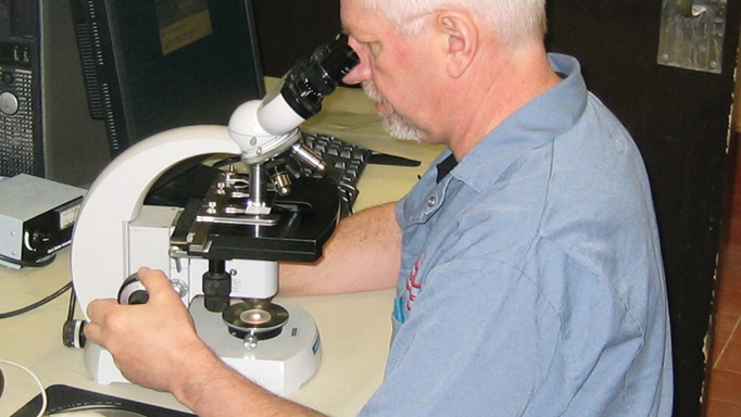
Following this process, Dr. Wojnarowicz decides which organs or parts should be taken for histopathology (examination under a microscope). The tissues are fixed in 10% buffered formalin solution for a few days and then trimmed and placed on histology slides which are read to establish a diagnosis. “Vets who do necropsies in the field will also send us fixed tissues for our diagnosis,” he adds. “But in winter, we have to have them fixed in alcohol rather than formalin, because the formalin will freeze in cold weather and then the slide becomes unreadable.” Depending on the horse’s history and Dr. Wojnarowicz’s findings so far, tissues may also be sent to other labs for testing for parasites, toxins, bacteria, or viruses.
Sometimes Dr. Wojnarowicz is sent horses who are suspected of having died of starvation because their owners could no longer feed them. He can detect starvation in two ways: by the way the normal fat around the horse’s heart and kidney becomes gel-like, and by changes in the bone marrow of the horse’s legs.
Performing a necropsy on a horse is a challenge, Dr. Wojnarowicz acknowledges. “Just the size and weight of the average horse makes it hard to do the dissections,” he says. “I am fortunate here to have good facility, equipment, and an excellent technologist to help. I take my hat off to those veterinarians in the field who do such a good job in difficult conditions.”

